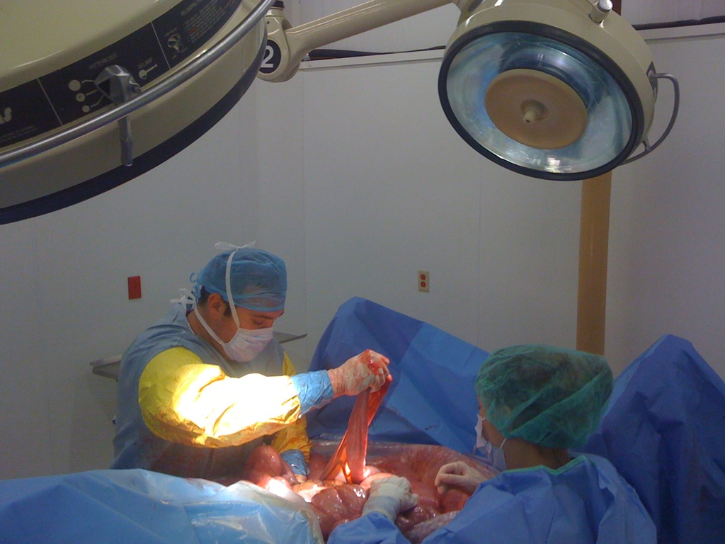
In a groundbreaking development in ocular health, CALEC surgery, or cultivated autologous limbal epithelial cell therapy, has emerged as a beacon of hope for patients suffering from severe corneal damage. Conducted at the renowned Mass Eye and Ear, this innovative procedure utilizes stem cells harvested from a healthy eye, leading to remarkable restoration of the corneal surface in patients with previously untreatable injuries. The trial demonstrated a striking success rate, with over 90 percent of participants achieving effective restoration of their corneas after receiving the treatment. With the ability to significantly enhance the quality of life for individuals facing chronic pain and vision impairment, CALEC surgery represents a pivotal advancement in corneal repair techniques. Through rigorous clinical trials, researchers are committed to ensuring this revolutionary method becomes widely available, paving the way for a brighter future in ocular therapeutic interventions.
The procedure known as cultivated autologous limbal epithelial cell therapy, or CALEC surgery, marks a transformative approach in restoring vision for those with corneal injuries. This surgical method has shown promising results by leveraging stem cell therapy to regenerate healthy ocular tissue, specifically limbal epithelial cells, which are essential for maintaining a transparent corneal surface. In clinical trials at Mass Eye and Ear, the technique proved successful in treating patients who had not previously benefited from traditional methods. With significant implications for ocular health, CALEC surgery could redefine treatment protocols for corneal repair and inspire further advancements in regenerative medicine. As researchers explore this innovative pathway, the hope is to expand treatment access and improve outcomes for patients suffering from corneal degenerative conditions.
Understanding CALEC Surgery: A Revolutionary Approach to Corneal Repair
Cultivated autologous limbal epithelial cell (CALEC) surgery is pioneering a new wave of therapeutic intervention for patients suffering from severe corneal damage. This innovative approach involves the transplantation of stem cells from a healthy eye to regenerate and repair the corneal surface in affected patients. Developed at Mass Eye and Ear, CALEC surgery has shown promise through clinical trials where it effectively restored corneal surfaces in a remarkable 90% of participants, illustrating its potential as a revolutionary treatment for what was previously considered untreatable eye injuries. This process not only highlights advancements in ocular health but also emphasizes the critical role that limbal epithelial cells play in maintaining the health of the eye, presenting new hope for those burdened by visual impairments due to corneal damage.
The CALEC procedure exemplifies the intersection of cutting-edge stem cell therapy and ophthalmic surgery by directly addressing the underlying causes of corneal health degradation. By extracting limbal epithelial cells from the patient’s healthy eye, these stem cells can be cultured into viable grafts before being meticulously transplanted into the damaged cornea. The implications of this surgical breakthrough extend beyond individual cases as it marks a substantial advancement in medical practices that could redefine standards for eye surgeries in the future.
Frequently Asked Questions
What is CALEC surgery and how does it relate to corneal repair?
CALEC surgery, which stands for cultivated autologous limbal epithelial cells, is an innovative procedure developed at Mass Eye and Ear for repairing corneal damage. It involves using stem cells sourced from a healthy eye to create a graft that restores the damaged cornea, offering new hope for patients with blinding corneal injuries.
How does stem cell therapy work in CALEC surgery?
In CALEC surgery, stem cell therapy involves harvesting limbal epithelial cells from a healthy eye through a biopsy. These cells are then expanded in a lab to form a cellular tissue graft. After a few weeks, this graft is transplanted into the damaged eye, effectively restoring the cornea’s surface and improving ocular health.
What are the success rates of CALEC surgery for corneal repair?
The clinical trial for CALEC surgery at Mass Eye and Ear reported high success rates, with nearly 90 percent of participants experiencing restoration of the corneal surface. Follow-ups indicated that 93 percent of patients achieved complete or partial success by 12 months post-surgery, highlighting the effectiveness of this stem cell procedure in treating corneal damage.
What are limbal epithelial cells and why are they important in CALEC surgery?
Limbal epithelial cells are healthy stem cells located at the limbus, the outer edge of the cornea. They play a crucial role in maintaining the eye’s smooth surface and optical clarity. In CALEC surgery, these cells are harvested to restore corneal integrity in patients who have suffered serious ocular injuries, showcasing their importance in treating corneal repair.
Is CALEC surgery available for patients at Mass Eye and Ear?
Currently, CALEC surgery remains experimental and is not widely available at Mass Eye and Ear or any U.S. hospitals. The procedure has undergone clinical trials, and further studies are needed before it can receive federal approval for wider application.
What are the potential risks or side effects of CALEC surgery?
While CALEC surgery has demonstrated a high safety profile in clinical trials, potential risks include minor adverse events such as infections or complications related to chronic contact lens use. Long-term studies will help further assess the safety of this innovative stem cell therapy.
What advancements are expected in CALEC surgery’s future?
Future advancements in CALEC surgery may include the development of an allogeneic manufacturing process using limbal stem cells from cadaveric donor eyes. This would expand the treatment’s applicability to patients with bilateral corneal damage, potentially improving ocular health for a broader patient population.
How can I find more information about CALEC surgery at Mass Eye and Ear?
For more information about CALEC surgery and ongoing clinical trials, you can visit the Mass Eye and Ear website or contact their Cornea Service directly. They provide updates on research developments and potential opportunities for patient participation in future study trials.
| Key Point | Details |
|---|---|
| First CALEC Surgery | Performed by Ula Jurkunas at Mass Eye and Ear. |
| Purpose of CALEC | To restore corneal surfaces using stem cells from healthy eyes. |
| Procedure Overview | Involves biopsy, cellular tissue graft creation (2-3 weeks), and surgical transplant. |
| Clinical Trial Success | 90% effective in restoring damaged corneal surfaces after 18 months. |
| Participants and Results | 14 patients, 50% fully restored at 3 months; 79% and 77% at 12 and 18 months respectively. |
| Safety Profile | High safety with no severe adverse events; minor issues resolved quickly. |
| Future Directions | Plans for broader application including allogeneic manufacturing for both eyes. |
Summary
CALEC surgery represents a groundbreaking advancement in the treatment of corneal injuries once deemed untreatable. By using stem cells to regenerate the cornea’s surface, this innovative procedure offers new hope for individuals suffering from vision impairments due to corneal damage. The promising results from clinical trials, demonstrating a high success rate and excellent safety profile, pave the way for further research and potential FDA approval. As such, CALEC surgery not only marks a significant milestone in ocular health but also opens doors for future therapeutic strategies in the field of ophthalmology.
